|
Small double-stranded
RNA (dsRNA) has been found to
silence gene expression by an evolutionally conserved mechanism known
as RNA interference or RNAi. Such dsRNAs are
called small interfering RNAs
or siRNA.
RNAi can occur at both transcriptional and post-transcriptional levels.
Surprisingly, various recent studies (see below) have found that dsRNA
can also activate
gene expression, a mechanism that has been termed "small
RNA-induced gene activation" or "saRNA" or "RNAa".
It has been shown that dsRNAs targeting gene promoters induce potent
transcriptional activation of associated genes. Both studies
demonstrate RNAa in human cells using synthetic dsRNAs termed small
activating RNAs (saRNAs). Endogenous microRNAs
(miRNA) that cause RNAa has also
been found in humans.
External links:
RNA interference: hitting the ON switch Researchers in San Francisco have findings that suggest a whole new side to RNA interference. Erika Check reports on their attempts to make a revolutionary field more revolutionary still. 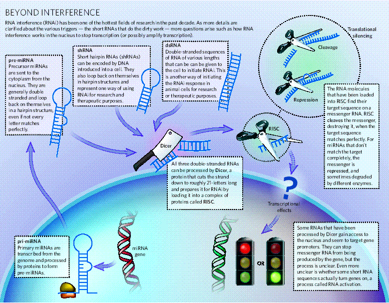 Turn genes on, turn diseases off Bob Holmes, New Scientist, 2007 http://www.science.org.au/nova/newscientist/098ns_001.htm  News and Views Q&A (by Helge Großhans and Witold Filipowicz) Molecular biology: The expanding world of small RNAs Molecular
cell biology has long been dominated
by a protein-centric view. But the emergence of small, non-coding RNAs
challenges this perception. These plentiful RNAs regulate gene
expression at different levels, and have essential roles in health and
disease.
When microRNAs acivate translation. Contrary to their
traditional role, microRNAs (miRNAs) contribute to an
increase in translation during cell quiescence. This function may be
exploited for microRNAmediated regulation of protein expression.
References: Small dsRNAs induce transcriptional activation in human cells. Li LC, Okino ST, Zhao H, Pookot D, Place RF, Urakami S, Enokida H, Dahiya R. Proc Natl Acad Sci U S A. 2006 103(46): 17337-17342 Department of Urology, Veterans Affairs Medical Center and University of California, San Francisco, CA 94121, USA. Recent studies have
shown that small noncoding RNAs, such as microRNAs and siRNAs, regulate
gene expression at multiple levels including chromatin architecture,
transcription, RNA editing, RNA stability, and translation. Each form
of RNA-dependent regulation has been generally found to silence
homologous sequences and collectively called RNAi. To further study the
regulatory role of small RNAs at the transcriptional level, we designed
and synthesized 21-nt dsRNAs targeting selected promoter regions of
human genes E-cadherin, p21(WAF1/CIP1) (p21), and VEGF. Surprisingly,
transfection of these dsRNAs into human cell lines caused long-lasting
and sequence-specific induction of targeted genes. dsRNA mutation
studies reveal that the 5' end of the antisense strand, or "seed"sequence,
is critical for activity. Mechanistically, the dsRNA-induced gene
activation requires the Argonaute 2 (Ago2) protein and is associated
with a loss of lysine-9 methylation on histone 3 at dsRNA-target sites.
In conclusion, we have identified several dsRNAs that activate gene
expression by targeting noncoding regulatory regions in gene promoters.
These findings reveal a more diverse role for small RNA molecules in
the regulation of gene expression than previously recognized and
identify a potential therapeutic use for dsRNA in targeted gene
activation.
PATENT: (WO/2006/113246) SMALL ACTIVATING RNA MOLECULES AND METHODS OF USE A small modulatory dsRNA specifies the fate of adult neural stem cells. Kuwabara T, Hsieh J, Nakashima K, Taira K, Gage FH. Cell. 2004 116(6): 779-793 Laboratory of Genetics, The Salk Institute, 10010 North Torrey Pines Road, La Jolla, CA 92037, USA. Discovering the molecular mechanisms that regulate neuron-specific gene expression remains a central challenge for CNS research. Here, we report that small, noncoding double-stranded (ds) RNAs play a critical role in mediating neuronal differentiation. The sequence defined by this dsRNA is NRSE/RE1, which is recognized by NRSF/REST, known primarily as a negative transcriptional regulator that restricts neuronal gene expression to neurons. The NRSE dsRNA can trigger gene expression of neuron-specific genes through interaction with NRSF/REST transcriptional machinery, resulting in the transition from neural stem cells with neuron-specific genes silenced by NRSF/REST into cells with neuronal identity that can express neuronal genes. The mechanism of action appears to be mediated through a dsRNA/protein interaction, rather than through siRNA or miRNA. The discovery of small modulatory dsRNAs (smRNAs) extends the important contribution of noncoding RNAs as key regulators of cell behavior at both transcriptional and posttranscriptional levels. Regulation of endothelial nitric oxide synthase by small RNA. Ming-Xiang Zhang *, Hesheng Ou *, Ying H. Shen, Jing Wang, Jian Wang, Joseph Coselli, and Xing Li Wang PNAS 2005 vol. 102 (47) 16967-16972 Division of Cardiothoracic Surgery, Michael E. DeBakey Department of Surgery, Baylor College of Medicine, and Texas Heart Institute at St Luke's Episcopal Hospital, Houston, TX 77030 Repeats (27-nt) in intron 4 have been shown to play a cis-acting role in endothelial nitric oxide synthase (eNOS) promoter activity. We hypothesize that the 27-nt repeats could be the source of small nuclear RNA specifically regulating eNOS expression. In this study, we used synthesized 27-nt RNA duplex and found that the eNOS gene transcriptional efficiency was reduced 63% (0.047 ± 0.009 vs. 0.126 ± 0.015, P < 0.01) by nuclear run-on assay. In endothelial cells transfected with the 27-nt small RNA duplex, we found that the eNOS mRNA and protein levels were decreased by >64% (P < 0.01). Conversely, a randomly selected 27-nt from luciferase gene had no effect on the eNOS expression. Furthermore, this eNOS silencing effect appeared to be reversible under the stimulation of vascular endothelial growth factor (10 ng/ml), which is known to up-regulate eNOS expression. Using in situ hybridization and Northern blotting, we observed the presence of endogenous eNOS intron 4-derived 27-nt small RNA, which was confined to the nucleus. In summary, we demonstrated that intron-based microRNAs in eNOS can induce significant gene specific transcriptional suppression, which could be an effective negative feedback regulator for gene expression. Activating gene expression in mammalian cells with promoter-targeted duplex RNAs. Janowski BA, Younger ST, Hardy DB, Ram R, Huffman KE, Corey DR. Nat Chem Biol. 2007 3(3): 166-173 Department of Pharmacology, University of Texas Southwestern Medical Center at Dallas, Dallas, Texas 75390, USA. The ability to selectively activate or inhibit gene expression is fundamental to understanding complex cellular systems and developing therapeutics. Recent studies have demonstrated that duplex RNAs complementary to promoters within chromosomal DNA are potent gene silencing agents in mammalian cells. Here we report that chromosome-targeted RNAs also activate gene expression. We have identified multiple duplex RNAs complementary to the progesterone receptor (PR) promoter that increase expression of PR protein and RNA after transfection into cultured T47D or MCF7 human breast cancer cells. Upregulation of PR protein reduced expression of the downstream gene encoding cyclooygenase 2 but did not change concentrations of estrogen receptor, which demonstrates that activating RNAs can predictably manipulate physiologically relevant cellular pathways. Activation decreased over time and was sequence specific. Chromatin immunoprecipitation assays indicated that activation is accompanied by reduced acetylation at histones H3K9 and H3K14 and by increased di- and trimethylation at histone H3K4. These data show that, like proteins, hormones and small molecules, small duplex RNAs interact at promoters and can activate or repress gene expression. Transcriptional activation by small RNA duplexes. John J. Rossi NATURE CHEMICAL BIOLOGY VOLUME 3 NUMBER 3 MARCH 2007 Division of Molecular Biology, Graduate School of Biological Sciences, Beckman Research Institute of the City of Hope, Duarte, California 91010, USA. Short double-stranded RNA duplexes are the triggers for post-transcriptional gene silencing and can also induce epigenetic silencing of genes at the level of transcription. A surprising new finding is that short RNA duplexes targeted to promoter regions can also mediate potent enhancement of transcription. Switching from repression to activation: microRNAs can up-regulate translation. Vasudevan S, Tong Y, Steitz JA. Science. 2007 318(5858): 1931-1934 Department of Molecular Biophysics and Biochemistry, Howard Hughes Medical Institute, Yale University School of Medicine, Boyer Center for Molecular Medicine, 295 Congress Avenue, New Haven, CT 06536, USA. AU-rich elements (AREs)
and microRNA target sites are conserved
sequences in messenger RNA (mRNA) 3' untranslated regions (3'UTRs) that
control gene expression posttranscriptionally. Upon cell cycle arrest,
the ARE in tumor necrosis factor-alpha (TNFalpha) mRNA is transformed
into a translation activation signal, recruiting Argonaute (AGO) and
fragile X mental retardation-related protein 1 (FXR1), factors
associated with micro-ribonucleoproteins (microRNPs). We show that
human microRNA miR369-3 directs association of these proteins with the
AREs to activate translation. Furthermore, we document that two
well-studied microRNAs-Let-7 and the synthetic microRNA
miRcxcr4-likewise induce translation up-regulation of target
mRNAs on cell cycle arrest, yet they repress translation in
proliferating cells. Thus, activation is a common function of microRNPs
on cell cycle arrest. We propose that translation regulation by
microRNPs oscillates between repression and activation during the cell
cycle.
Activation of an oncogenic microRNA cistron by provirus integration. Wang CL, Wang BB, Bartha G, Li L, Channa N, Klinger M, Killeen N, Wabl M. Proc Natl Acad Sci U S A. 2006 103(49): 18680-18684 Department of Microbiology and Immunology, University of California-San Francisco, San Francisco, CA 94143, USA. Retroviruses can cause
tumors when they integrate near a protooncogene
or tumor suppressor gene of the host. We infected >2,500 mice with
the SL3-3 murine leukemia virus; in 22 resulting tumors, we found
provirus integrations nearby or within the gene that contains the
mir-17-92 microRNA (miRNA) cistron. Using quantitative real-time PCR,
we showed that expression of miRNA was increased in these tumors,
indicating that retroviral infection can induce expression of oncogenic
miRNAs. Our results demonstrate that retroviral mutagenesis can be a
potent tool for miRNA discovery.
MicroRNA-373 induces expression of genes with complementary promoter sequences. Robert F. Place, Long-Cheng Li, Deepa Pookot, Emily J. Noonan, and Rajvir Dahiya PNAS (2008) 105(5) 1608-1613 Recent studies have
shown that microRNA (miRNA) regulates gene expression by repressing
translation or directing sequence-specific degradation of complementary
mRNA. Here, we report new evidence in which miRNA may also function to
induce gene expression. By scanning gene promoters in silico for
sequences complementary to known miRNAs, we identified a putative
miR-373 target site in the promoter of E-cadherin. Transfection of
miR-373 and its precursor hairpin RNA (pre-miR-373) into PC-3 cells
readily induced E-cadherin expression. Knockdown experiments confirmed
that induction of E-cadherin by pre-miR-373 required the miRNA
maturation protein Dicer. Further analysis revealed that cold-shock
domain-containing protein C2 (CSDC2), which possesses a putative
miR-373 target site within its promoter, was also readily induced in
response to miR-373 and pre-miR-373. Furthermore, enrichment of RNA
polymerase II was detected at both E-cadherin and CSDC2 promoters after
miR-373 transfection. Mismatch mutations to miR-373 indicated that gene
induction was specific to the miR-373 sequence. Transfection of
promoter-specific dsRNAs revealed that the concurrent induction of
E-cadherin and CSDC2 by miR-373 required the miRNA target sites in both
promoters. In conclusion, we have identified a miRNA that targets
promoter sequences and induces gene expression. These findings reveal a
new mode by which miRNAs may regulate gene expression.
Silencing of microRNAs in vivo with 'antagomirs'. Krützfeldt J, Rajewsky N, Braich R, Rajeev KG, Tuschl T, Manoharan M, Stoffel M. Nature. 2005 438(7068): 685-689 Laboratory of Metabolic Diseases, The Rockefeller University, 1230 York Avenue, New York, New York 10021, USA. MicroRNAs (miRNAs) are
an abundant class of non-coding RNAs that are believed to be important
in many biological processes through regulation of gene expression. The
precise molecular function of miRNAs in mammals is largely unknown and
a better understanding will require loss-of-function studies in vivo.
Here we show that a novel class of chemically engineered
oligonucleotides, termed 'antagomirs', are efficient and specific
silencers of endogenous miRNAs in mice. Intravenous administration of
antagomirs against miR-16, miR-122, miR-192 and miR-194 resulted in a
marked reduction of corresponding miRNA levels in liver, lung, kidney,
heart, intestine, fat, skin, bone marrow, muscle, ovaries and adrenals.
The silencing of endogenous miRNAs by this novel method is specific,
efficient and long-lasting. The biological significance of silencing
miRNAs with the use of antagomirs was studied for miR-122, an abundant
liver-specific miRNA.Gene expression and bioinformatic analysis of
messenger RNA from antagomir-treated animals revealed that the 3'
untranslated regions of upregulated genes are strongly
enriched in miR-122 recognition motifs, whereas downregulated genes are
depleted in these motifs. Analysis of the functional annotation of
downregulated genes specifically predicted that cholesterol
biosynthesis genes would be affected by miR-122, and plasma cholesterol
measurements showed reduced levels in antagomir-122-treated mice. Our
findings show that antagomirs are powerful tools to silence specific
miRNAs in vivo and may represent a therapeutic strategy for silencing
miRNAs in disease.
Specificity, duplex degradation and subcellular localization of antagomirs. Krützfeldt J, Kuwajima S, Braich R, Rajeev KG, Pena J, Tuschl T, Manoharan M, Stoffel M. Nucleic Acids Res. 2007;35(9): 2885-2892 Laboratory of Metabolic Diseases, The Rockefeller University, 1230 York Avenue, New York, NY 10021, USA. MicroRNAs (miRNAs) are
an abundant class of 20-23-nt long regulators of gene expression. The
study of miRNA function in mice and potential therapeutic approaches
largely depend on modified oligonucleotides. We recently demonstrated
silencing miRNA function in mice using chemically modified and
cholesterol-conjugated RNAs termed 'antagomirs'. Here, we further
characterize the properties and function of antagomirs in mice. We
demonstrate that antagomirs harbor optimized phosphorothioate
modifications, require >19-nt length for highest efficiency and can
discriminate between single nucleotide mismatches of the targeted
miRNA. Degradation of different chemically protected miRNA/antagomir
duplexes in mouse livers and localization of antagomirs in a cytosolic
compartment that is distinct from processing (P)-bodies indicates a
degradation mechanism independent of the RNA interference (RNAi)
pathway. Finally, we show that antagomirs, although
incapable of silencing miRNAs in the central nervous system (CNS) when
injected systemically, efficiently target miRNAs when injected locally
into the mouse cortex. Our data further validate the effectiveness of
antagomirs in vivo and should facilitate future studies to silence
miRNAs for functional analysis and in clinically relevant settings.
Small RNA-mediated Gene Activation EpiGenie recently reviewed the epigenetic text RNA and the Regulation of Gene Expression by Kevin Morris. To give you a small taste of the material covered in the book, here is a summary of one of the chapters: Small RNA-mediated Gene Activation by Long-Cheng Li (2008) Noncoding RNAs (ncRNA) which, by definition, do not code for proteins, are estimated to make up ~98% of the human transcriptome. Once thought to be the transcriptional by-product of “junk DNA,” ncRNA is rapidly becoming recognized as an important mechanism of gene regulation. Small double-stranded RNAs (dsRNA) in particular have been extensively studied in the context of gene silencing by a mechanism commonly referred to as RNA interference (RNAi). This chapter focuses on the lesser known flip-side of RNAi, which involves small dsRNA-mediated gene activation (RNAa). Known functions of ncRNA Noncoding RNA can be divided into 2 major functional classes: 1) housekeeping ncRNAs and 2) regulatory ncRNAs. Well-studied housekeeping functions include those involved in RNA splicing, editing, and protein synthesis. More recently discovered housekeeping functions of ncRNA involve serving as reaction catalysts (ribozymes) and molecular sensors (riboswitches). The regulatory functions of ncRNA, on the other hand, directly impact gene expression through a variety of mechanisms. While large regulatory RNA (hundreds to thousands of nucleotides long) are known to participate in diverse processes such as dosage compensation and imprinting, the focus of this chapter is on small regulatory dsRNA with a size range of 20-30 nucleotides. Small dsRNAs as negative regulators of gene expression RNA interference by exogenous dsRNA was first reported in Caenorhabditis elegans in 1998. Subsequent studies showed that small endogenous dsRNA, called microRNA (miRNA), also participate in RNAi in a trans-acting, target sequence-specific manner similar to that reported for exogenously introduced small interfering RNA (siRNA). While the mechanism of miRNA-mediated gene suppression has now been elaborated with respect to the proteins involved and RNA processing, the relationship between RNAi and long-term suppression of gene expression through RNA-directed DNA methylation (which has been observed in plants) or other epigenetic mechanisms is still unknown. dsRNA-induced transcriptional activation In contrast to RNAi, RNA activation (RNAa) mediated by small activating RNAs (saRNAs) is a relatively recent (2006/2007) discovery. Like RNAi, RNAa targets specific genes by sequence complementarity, with target specificity determined primarily by the 5’-end of the saRNA molecule. However, the time course of RNAa is very different from that of RNAi, with induction of gene expression occurring 2-3 days post-transfection as opposed to several hours for RNAi-mediated suppression, and the effects of RNAa appear to be longer lasting (at least 10-15 days vs. 5-7 days for RNAi). The reasons for these differences in time course are not clear, but it has been postulated (based on some evidence) that the slow induction reflects the time required for saRNA to gain access to DNA (possibly occurring only during mitosis), and that the prolonged effects on gene expression may be due to longer-lasting RNAa-induced epigenetic changes in chromatin. Given that expression of rare transcripts can be increased 5- to 10-fold, RNAa can be a potent mechanism for altering gene expression and restoring function of silenced genes. miRNAs and bidirectional gene regulation MicroRNAs are small endogenous ncRNA that are produced by the elaborate processing of longer precursor RNA molecules. While much of the attention regarding miRNA has focused on its role in the inhibition of gene expression, the activation of gene expression in response to miRNA has been generally overlooked as a secondary effect. In light of what recent studies on saRNA have shown, it would be of utmost importance to further investigate the potential role of miRNA in gene activation. Future of small RNA Gene Activation The recent discovery of RNA activation by small dsRNA raises the exciting possibility that genes silenced by disease might be turned on again in a gene-specific manner by saRNA. However, much more work needs to be done to first understand the mechanism and limitations of dsRNA-mediated gene activation before saRNA can be applied therapeutically. Together, the phenomena of RNAa and RNAi demonstrate the complexity as well as yin-yang nature of gene regulation by small double-stranded ncRNA. RNAi and RNAa -- the yin and yang of RNAome. Pushparaj PN, Aarthi JJ, Kumar SD, Manikandan J. Department of Physiology, Yong Loo Lin School of Medicine, National University of Singapore, Singapore. phspnp@nus.edu.sg Bioinformation. 2008 Jan 11;2(6): 235-237 RNA interference (RNAi) is a powerful technology with huge applications for functional genomics, target identification in drug discovery and elucidation of molecular signaling pathways. Current RNAi studies have demonstrated the clinical potential of small interfering RNAs (siRNAs) in metabolic diseases, cancer, AIDS, malaria, neurodegenerative disorders, dental diseases and other illnesses. Interestingly, recent studies have shown that the small RNA molecules, either indigenously produced as microRNAs (miRNAs) or exogenously administered synthetic dsRNAs could effectively activate a particular gene in a sequence specific manner instead of silencing it. This novel, but still uncharacterized, phenomenon has been termed as RNA activation (RNAa). The paradoxical concept of Yin and Yang, which describe two primal opposing but complementary principles, can potentially be applied to elucidate the complex phenomenon of RNAa/RNAi in the RNAome. This warrants a proper understanding of the RNAi/RNAa molecular pathways in living organisms before any of the small dsRNAs can potentially be exploited for therapeutics in human beings. Novel functions for small RNA molecules. Zhang C. University of Medicine and Dentistry of New Jersey, New Jersey Medical School, RNA Research Laboratory, Department of Anesthesiology, 185 South Orange Avenue, MSB Room E548, Newark, NJ 07101, USA Curr Opin Mol Ther. 2009 Dec;11(6): 641-651. Small RNAs are short (approximately 18 to 30 nucleotides), non-coding RNA molecules that can regulate gene expression in both the cytoplasm and the nucleus via post-transcriptional gene silencing (PTGS), chromatin-dependent gene silencing (CDGS) or RNA activation (RNAa). Three classes of small RNAs have been defined: microRNAs (miRNAs), siRNAs and Piwi-interacting RNAs (piRNAs). Research has indicated that small RNAs play important roles in cellular processes such as cell differentiation, growth/proliferation, migration, apoptosis/death, metabolism and defense. Accordingly, small RNAs are critical regulators of normal development and physiology. More interestingly, increasing evidence indicates that small RNAs are involved in the pathogenesis of diverse diseases including cancer, cardiovascular disease, stroke, neurodegenerative disease, diabetes, liver disease, kidney disease and infectious disease. More than 20 clinical trials are ongoing to evaluate therapies based on small RNA. Additionally, small RNAs may serve as novel biomarkers and therapeutic targets for the majority of diseases. siRNA, miRNA, and shRNA: in vivo applications. Pushparaj PN, Aarthi JJ, Manikandan J, Kumar SD. Department of Physiology, National University of Singapore, Singapore J Dent Res. 2008 Nov;87(11): 992-1003. RNA interference (RNAi), an accurate and potent gene-silencing method, was first experimentally documented in 1998 in Caenorhabditis elegans by Fire et al., who subsequently were awarded the 2006 Nobel Prize in Physiology/Medicine. Subsequent RNAi studies have demonstrated the clinical potential of synthetic small interfering RNAs (siRNAs) or short hairpin RNAs (shRNAs) in dental diseases, eye diseases, cancer, metabolic diseases, neurodegenerative disorders, and other illnesses. siRNAs are generally from 21 to 25 base-pairs (bp) in length and have sequence-homology-driven gene-knockdown capability. RNAi offers researchers an effortless tool for investigating biological systems by selectively silencing genes. Key technical aspects--such as optimization of selectivity, stability, in vivo delivery, efficacy, and safety--need to be investigated before RNAi can become a successful therapeutic strategy. Nevertheless, this area shows a huge potential for the pharmaceutical industry around the globe. Interestingly, recent studies have shown that the small RNA molecules, either indigenously produced as microRNAs (miRNAs) or exogenously administered synthetic dsRNAs, could effectively activate a particular gene in a sequence-specific manner instead of silencing it. This novel, but still uncharacterized, phenomenon has been termed 'RNA activation' (RNAa). In this review, we analyze these research findings and discussed the in vivo applications of siRNAs, miRNAs, and shRNAs. RNAa is conserved in mammalian cells. Huang V, Qin Y, Wang J, Wang X, Place RF, Lin G, Lue TF, Li LC. Helen-Diller Comprehensive Cancer Center, University of California San Francisco, San Francisco, California, USA. PLoS One. 2010 Jan 22;5(1): e8848 BACKGROUND: RNA activation (RNAa) is a newly discovered mechanism of gene activation triggered by small double-stranded RNAs termed 'small activating RNAs' (saRNAs). Thus far, RNAa has only been demonstrated in human cells and is unclear whether it is conserved in other mammals. METHODOLOGY/PRINCIPAL FINDINGS: In the present study, we evaluated RNAa in cells derived from four mammalian species including nonhuman primates (African green monkey and chimpanzee), mouse, and rat. Previously, we identified saRNAs leading to the activation of E-cadherin, p21, and VEGF in human cells. As the targeted sequences are highly conserved in primates, transfection of each human saRNA into African green monkey (COS1) and chimpanzee (WES) cells also resulted in induction of the intended gene. Additional saRNAs targeting clinically relevant genes including p53, PAR4, WT1, RB1, p27, NKX3-1, VDR, IL2, and pS2 were also designed and transfected into COS1 and WES cells. Of the nine genes, p53, PAR4, WT1, and NKX3-1 were induced by their corresponding saRNAs. We further extended our analysis of RNAa into rodent cell types. We identified two saRNAs that induced the expression of mouse Cyclin B1 in NIH/3T3 and TRAMP C1 cells, which led to increased phosphorylation of histone H3, a downstream marker for chromosome condensation and entry into mitosis. We also identified two saRNAs that activated the expression of CXCR4 in primary rat adipose-derived stem cells. CONCLUSIONS/SIGNIFICANCE: This study demonstrates that RNAa exists in mammalian species other than human. Our findings also suggest that nonhuman primate disease models may have clinical applicability for validating RNAa-based drugs. Small RNA and transcriptional upregulation. Portnoy V, Huang V, Place RF, Li LC. Department of Urology and Helen-Diller Comprehensive Cancer Center, University of California San Francisco, USA. Wiley Interdiscip Rev RNA. 2011 Sep-Oct;2(5): 748-60 Small RNA molecules, such as microRNA and small interfering RNA, have emerged as master regulators of gene expression through their ability to suppress target genes in a phenomenon collectively called RNA interference (RNAi). There is growing evidence that small RNAs can also serve as activators of gene expression by targeting gene regulatory sequences. This novel mechanism, known as RNA activation (RNAa), appears to be conserved in at least mammalian cells and triggered by both endogenous and artificially designed small RNAs. RNAa depends on Argonaute proteins, but possesses kinetics distinct from that of RNAi. Epigenetic changes are associated with RNAa and may contribute to transcriptional activation of target genes, but the underlying mechanism remains elusive. Given the potential of RNAa as a molecular tool for studying gene function and as a therapeutic for disease, further research is needed to completely elucidate its molecular mechanism in order to refine the rules for target selection and improve strategies for exploiting it therapeutically. WIREs RNA 2011 2 748-760 DOI: 10.1002/wrna.90 For further resources related to this article, please visit the WIREs website. Upregulation of Cyclin B1 by miRNA and its implications in cancer. Vera Huang, Robert F. Place, Victoria Portnoy, Ji Wang, Zhongxia Qi, Zhejun Jia, Angela Yu, Marc Shuman, Jingwei Yu, and Long-Cheng Li Nucleic Acids Res. 2012 February; 40(4): 1695–1707 It is largely recognized that microRNAs (miRNAs) function to silence gene expression by targeting 3′UTR regions. However, miRNAs have also been implicated to positively-regulate gene expression by targeting promoter elements, a phenomenon known as RNA activation (RNAa). In the present study, we show that expression of mouse Cyclin B1 (Ccnb1) is dependent on key factors involved in miRNA biogenesis and function (i.e. Dicer, Drosha, Ago1 and Ago2). In silico analysis identifies highly-complementary sites for 21 miRNAs in the Ccnb1 promoter. Experimental validation identified three miRNAs (miR-744, miR-1186 and miR-466d-3p) that induce Ccnb1 expression in mouse cell lines. Conversely, knockdown of endogenous miR-744 led to decreased Ccnb1 levels. Chromatin immunoprecipitation (ChIP) analysis revealed that Ago1 was selectively associated with the Ccnb1 promoter and miR-744 increased enrichment of RNA polymerase II (RNAP II) and trimethylation of histone 3 at lysine 4 (H3K4me3) at the Ccnb1 transcription start site. Functionally, short-term overexpression of miR-744 and miR-1186 resulted in enhanced cell proliferation, while prolonged expression caused chromosomal instability and in vivo tumor suppression. Such phenotypes were recapitulated by overexpression of Ccnb1. Our findings reveal an endogenous system by which miRNA functions to activate Ccnb1 expression in mouse cells and manipulate in vivo tumor development/growth. Targeted induction of endogenous NKX3-1 by small activating RNA inhibits prostate tumor growth. Ren S, Kang MR, Wang J, Huang V, Place RF, Sun Y, Li LC. Department of Urology and Helen-Diller Comprehensive Cancer Center, University of California San Francisco, San Francisco, California; Department of Urology, Shanghai Changhai Hospital, Second Military Medical University, Shanghai, China. Prostate. 2013 Jul 8 [Epub ahead of print] BACKGROUND: RNA activation (RNAa) is a small RNA-mediated gene regulation mechanism by which expression of a particular gene can be induced by targeting its promoter using small double-stranded RNA also known as small activating RNA (saRNA). We used saRNA as a molecular tool to examine NKX3-1's role as a tumor suppressor and tested in vitro and in vivo antitumor effects of NKX3-1 induction by saRNA. MATERIALS AND METHODS: NKX3-1 saRNA was transfected into human prostate cancer cells including LNCaP, CWR22R, PC-3, CWR22RV1, DuPro, LAPC4, and DU145. The transfected cells were used for analysis of gene expression by RT-PCR and immunoblotting, proliferation, apoptosis and cell cycle distribution. PC-3 xenograft models were established in immunocompromised mice and treated with NKX3-1 saRNA. RESULTS: NKX3-1 saRNA induced NKX3-1 expression in different prostate cancer cell lines, resulting in inhibited cell proliferation and survival, cell cycle arrest and apoptotic cell death. These effects were partly mediated by NKX3-1's regulation of several downstream genes including the upregulation of p21 and p27, and the inhibition of VEGFC expression. Treatment of mouse xenograft prostate tumors with intratumoral delivery of NKX3-1 saRNA formulated in lipid nanoparticles significantly inhibited tumor growth and prolonged animal survival. CONCLUSIONS: By revealing several important target genes of NKX3-1, our findings corroborated NKX3-1's role as a tumor suppressor gene through direct regulation of the cell cycle and growth/survival pathways. This study also validated the therapeutic potential of saRNA for the treatment of prostate cancer via targeted activation of tumor suppressor genes. Involvement of argonaute proteins in gene silencing and activation by RNAs complementary to a non-coding transcript at the progesterone receptor promoter. Yongjun Chu, Xuan Yue, Scott T. Younger, Bethany A. Janowski, and David R. Corey Nucleic Acids Res. 2010 November; 38(21): 7736–7748. Double-stranded RNAs that are complementary to non-coding transcripts at gene promoters can activate or inhibit gene expression in mammalian cells. Understanding the mechanism for modulating gene expression by promoter-targeted antigene RNAs (agRNAs) will require identification of the proteins involved in recognition. Previous reports have implicated argonaute (AGO) proteins, but identifications have differed with involvement of AGO1, AGO2, or both AGO1 and AGO2 being reported by different studies. The roles of AGO3 and AGO4 have not been investigated. Here, we examine the role of AGO 1–4 in gene silencing and activation of the progesterone receptor (PR) gene. Expression of AGO2 is necessary for efficient gene silencing or activation and AGO2 is recruited to the non-coding transcript that overlaps the promoter during both gene silencing and activation. Expression of AGO1, AGO3 and AGO4 are not necessary for gene silencing or activation nor are AGO1, AGO3, or AGO4 recruited to the target non-coding transcript during gene activation. These data indicate that AGO2 is the primary AGO variant involved in modulating expression of PR by agRNAs. Small RNA and its application in andrology and urology. Wang J, Li LC. Department of Urology and Helen-Diller Comprehensive Cancer Center, University of California San Francisco, San Francisco, CA 94158, USA. Transl Androl Urol. 2012;1(1): 33-43. Small non-coding RNAs such as small interfering RNA (siRNA), microRNA (miRNA) and piwi-interacting RNA (piRNA) exist in almost all kingdoms of organisms and have recently emerged as master regulators of gene expression to affect a diverse range of important biological processes. They exert their functions largely through two related but opposing mechanisms: RNA interference (RNAi) mediated by siRNA, miRNA and piRNA, and RNA activation (RNAa) mediated by small activating RNA (saRNA) and miRNA, leading to silencing and overexpression of target genes respectively. Dysregulation of these mechanisms have been implicated in a variety of human diseases including urological and andrological diseases. Importantly, both mechanisms can be readily harnessed for therapeutic purposes for a variety of diseases by using small RNA molecules as the "ribodrug". In this review, we highlight recent advances in the applications of small RNA as therapeutics for urological cancer, male infertile and erectile dysfunction. Defining features and exploring chemical modifications to manipulate RNAa activity. Place RF, Noonan EJ, Földes-Papp Z, Li LC. Helen Diller Comprehensive Cancer Center, Department of Urology, University of California, PO Box 589001, San Francisco, CA 94158-9001, USA Curr Pharm Biotechnol. 2010 Aug;11(5): 518-526 RNA interference (RNAi) is an evolutionary conserved mechanism by which small double-stranded RNA (dsRNA)--termed small interfering RNA (siRNA)--inhibit translation or degrade complementary mRNA sequences. Identifying features and enzymatic components of the RNAi pathway have led to the design of highly-effective siRNA molecules for laboratory and therapeutic application. RNA activation (RNAa) is a newly discovered mechanism of gene induction also triggered by dsRNAs termed small activating RNA (saRNA). It offers similar benefits as RNA interference (RNAi), while representing a new method of gene overexpression. In the present study, we identify features of RNAa and explore chemical modifications to saRNAs that improve the applicability of RNAa. We evaluate the rate of RNAa activity in order to define an optimal window of gene induction, while comparing the kinetic differences between RNAa and RNAi. We identify Ago2 as a conserved enzymatic component of both RNAa and RNAi implicating that saRNA may tolerate modification based on Ago2 function. As such, we define chemical modifications to saRNAs that manipulate RNAa activity, as well as exploit their effects to design saRNAs with enhanced medicinal properties. These findings reveal functional features of RNAa that may be utilized to augment saRNA function for mechanistic studies or the development of RNAa-based drugs. Up-regulation of VEGF by small activator RNA in human corpus cavernosum smooth muscle cells. Chen R, Wang T, Rao K, Yang J, Zhang S, Wang S, Liu J, Ye Z. Department of Urology, Tongji Hospital, Tongji Medical College, Huazhong University of Science and Technology, Wuhan, Hubei, China. J Sex Med. 2011 Oct;8(10): 2773-2780 INTRODUCTION: Functional failure of smooth muscle cells and endothelial cells in corpus cavernosum contributes to erectile dysfunction (ED) in aging men. Given that vascular endothelial growth factor (VEGF) may improve the function of smooth muscle cells and endothelial cells through different mechanisms, it is thus expected that increasing the expression of VEGF may have beneficial effects on erectile function. AIM: The aim of this article is to explore the possibility that VEGF can be induced by ribonucleic acid activation (RNAa) technology, and VEGF induction by RNAa has the potential of treating ED. METHODS: Primary human corpus cavernosum smooth muscle cells (CCSMCs) were isolated and cultured in vitro. The expression of α-smooth muscle actin was detected by immunohistochemistry to identify CCSMCs. A previously identified VEGF promoter-targeted small activator RNA (saRNA, double-stranded [ds]VEGF-706) and a negative control dsRNA were chemically synthesized. Cultured human CCSMCs were transfected with the saRNAs. The expression of VEGF messenger RNA (mRNA) and protein in transfected CCSMCs was evaluated by real-time polymerase chain reaction (RT-PCR) and Western blotting assay, respectively. Immunofluorescent staining was also used to confirm VEGF protein expression in cultured CCSMCs. MAIN OUTCOME MEASURE: The expression of VEGF was assessed by RT quantitative PCR, Western blotting, and immunofluorescence assays. RESULTS: After transfection, RT quantitative PCR analysis showed that the expression of VEGF mRNA was significantly induced in dsVEGF-706 transfected cells compared with cells receiving control treatments (P < 0.05). Consistent with mRNA induction, Western blotting and immunofluorescence analysis showed that VEGF protein expression was also induced by dsVEGF-706. CONCLUSION: VEGF expression can be activated by RNAa in primary human CCSMCs, suggesting a potential application of RNAa-mediated VEGF activation for the treatment of ED. RNA and the Regulation of Gene Expression - A Hidden Layer of Complexity Publisher: Caister Academic Press Edited by: Kevin V. Morris The Scripps Research Institute, La Jolla, USA Publication date: March 2008 ISBN: 978-1-904455-25-7 The role of RNA in regulating gene expression has become a topic of intense interest. In this book internationally recognized experts in RNA research explore and discuss the methods whereby RNA can regulate gene expression with examples in yeast, Drosophila, mammals, and viral infection, and highlight the application of this knowledge in therapeutics and research. Topics include: gene silencing and gene activation, the hammerhead ribozyme, epigenetic regulation, RNAi, microRNA, and pyknons. This comprehensive publication is intended for readers with teaching or research interests in RNA, the regulation of gene expression, genetics, genomics or molecular biology. Reviews: "the contributions in this book do provide informative and well-structured overviews of current understanding of the roles of non-coding RNAs, short interfering RNAs, microRNAs and retrotransposons in eukaryotic organisms ... cutting edge studies on the potential role of RNA species in the epigenetic regulation of gene expression and on the existence of previously unidentified classes of intergenic and intronic short regulatory RNAs (pyknons) ... a useful purchase for specialist workers in the field as well as for many institutional libraries." from Microbiology Today (2008) "This book is a well-selected compilation of 14 mostly review-style articles, written by experts in the field ... a well-written, successful endeavour to present the field of eukaryal RNA-mediated regulation of gene expression. It has its major strength in providing an extremely well structured, up-to-date, comprehensive overview that skillfully zooms the reader into each topic from a general introduction to a high degree of detail ... suited for a broad range of readers, from advanced students to researchers in the field. Personally, we very much enjoyed reading it." from ChemBioChem (2008) 9: 2005-2007 Content: Chapter 1 - The Hammerhead Ribozyme Revisited: New Biological Insights for the Development of Therapeutic Agents and for Reverse Genomics Applications Justin Hean and Marc S. Weinberg Chapter 2 - Epigenetic Regulation of Gene Expression Kevin V. Morris Chapter 3 - The Role of RNAi and Noncoding RNAs in Polycomb Mediated Control of Gene Expression and Genomic Programming Manuela Portoso and Giacomo Cavalli Chapter 4 - Heterochromatin Assembly and Transcriptional Gene Silencing under the Control of Nuclear RNAi: Lessons from Fission Yeast Aurélia Vavasseur, Leila Touat-Todeschini and André Verdel Chapter 5 - RNA-Mediated Gene Regulation in Drosophila Harsh H. Kavi, Harvey R. Fernandez, Weiwu Xie and James A. Birchler Chapter 6 - MicroRNA-Mediated Regulation of Gene Expression Lena J. Chin and Frank J. Slack Chapter 7 - Viral Infection-Related MicroRNAs in Viral and Host Genomic Evolution Yoichi R. Fujii and Nitin K. Saksena Chapter 8 - Regulation of Mammalian Mobile DNA by RNA-Based Silencing Pathways Harris Soifer Chapter 9 - The Role of Non-Coding RNAs in Controlling Mammalian RNA Polymerase II Transcription Stacey D. Wagner, Jennifer F. Kugel and James A. Goodrich Chapter 10 - Pyknons as Putative Novel and Organism-Specific Regulatory Motifs Isidore Rigoutsos Chapter 11 - RNA-Mediated Recognition of Chromosomal DNA David R. Corey Chapter 12 - RNA Mediated Transcriptional Gene Silencing: Mechanism and Implications in Writing the Histone Code Kevin V. Morris Chapter 13 - Small RNA-Mediated Gene Activation Long-Cheng Li Chapter 14 - Therapeutic Potential of RNA-mediated Control of Gene Expression: Options and Designs Lisa Scherer and John J. Rossi |
